
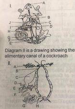
Diagram 1 is a drawing showing the alimentary canal of a mammal. Study the diagram carefully (a) Name the structure labelled A to J (b) What are the functions of the parts labelled A, C and H?
(C) Name the parts labelled A to H (d) State the functions of the parts labelled D and E (e) State two features common diagram I and D in diagram II (f) identify and name the parts in diagrams I and ll that perform similar functions
Explanation
(a) The names of the structures labelled A to J are A = Teeth (tooth) or incisors (incisor), B = Oesophagus or gullet C = Stomach D = Duodenum E = Rectum, F =Appendix, G = Large intestine or caecum., H= Smal intestine, I = Bile duct or gall bladder, J=Liver.
(b) The functions of the part labelled A, C and H are: (i) A = for biting or piercing or gnawing or cutting food into small pieces (ii) C= for (temporary) storage or for further digestion of food H= for digestion or absorption of food (c) The parts labelled A to H in diagram ii are: A= Salivary gland, B = Oesophagus or gullet C= Crop D= Gizzard E= Hepatic caeca or digestive gland or enteric caeca or mesenteric caeca F = Mid-gut or middle gut or mesenteron, G= small intestine or Hind gut H = Rectum (d) The functions of the parts labelled D and E are: D = for grinding or crushing food Common to the parts labelled C in diagram I and D in diagram II are: Both are (i) muscular and (ii) have sac-like or convoluted or rough interior (f) The parts in diagrams I and l that perform similar Functions are: Salivary glands, oesophagus, crop or stomach, pancreas or hepatic caeca, ileum or small intestine, large intestine or colon or rectum and anus.