ANWSER
Question 1:
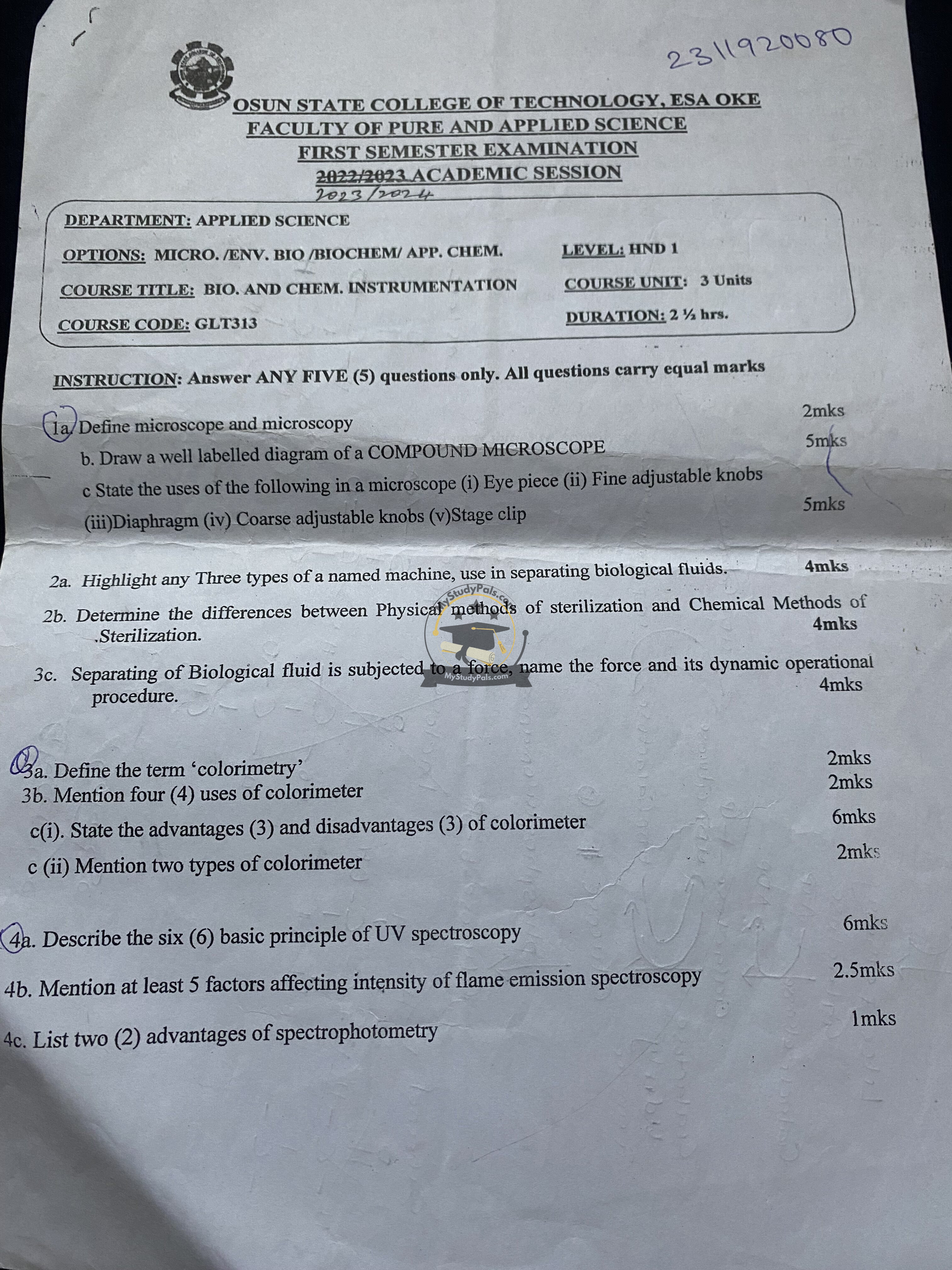
(a) Define microscope and microscopy
- A microscope is an optical instrument used to magnify small objects, allowing them to be seen in detail.
- Microscopy is the science of using microscopes to view objects that are too small to be seen by the naked eye.
(b) Draw a well-labelled diagram of a compound microscope
(Diagram should be drawn showing key parts: eyepiece, objective lens, stage, coarse and fine adjustment knobs, diaphragm, and light source.)
(c) State the uses of the following in a microscope:
- Eyepiece: Magnifies the image formed by the objective lens.
- Fine adjustable knobs: Used for precise focusing to get a sharp image.
- Diaphragm: Regulates the amount of light entering the microscope.
- Coarse adjustable knobs: Moves the stage up and down for initial focusing.
- Stage clip: Holds the slide in place on the stage.
Question 2:
(a) Highlight any three types of a named machine used in separating biological fluids.
- Centrifuge – Separates fluids based on density using centrifugal force.
- Dialysis Machine – Used for filtering waste from blood in patients with kidney failure.
- Chromatography Machine – Separates components of biological mixtures based on solubility.
(b) Determine the differences between physical and chemical methods of sterilization.
| Physical Sterilization | Chemical Sterilization |
|---|---|
| Uses heat, radiation, or filtration. | Uses chemical agents like ethylene oxide, alcohol, and formaldehyde. |
| Examples: Autoclave, UV sterilization, filtration. | Examples: Alcohol wipes, hydrogen peroxide, bleach solutions. |
| Physical damage to microorganisms. | Chemical reactions kill microorganisms. |
(c) Separating biological fluid is subjected to a force, name the force and its dynamic operational procedure.
- Force: Centrifugal Force
- Operational Procedure: A centrifuge spins biological samples at high speed, creating a force that separates components based on density (e.g., blood separation into plasma and cells).
Question 3:
(a) Define the term “colorimetry”.
- Colorimetry is the science of measuring the absorption of light at a specific wavelength by a colored solution to determine the concentration of a substance.
(b) Mention four (4) uses of a colorimeter.
- Determining concentration of colored solutions.
- Measuring enzyme activity in biochemical analysis.
- Water and food quality testing.
- Clinical diagnostics, e.g., hemoglobin measurement.
(c)(i) State three (3) advantages and three (3) disadvantages of a colorimeter.
Advantages:
- Simple and easy to use.
- Provides quick and accurate results.
- Cost-effective compared to spectrophotometers.
Disadvantages:
- Limited to colored solutions.
- Less sensitive than advanced spectrophotometers.
- Cannot measure absorbance in ultraviolet or infrared ranges.
(c)(ii) Mention two types of colorimeter.
- Visual Colorimeter
- Photoelectric Colorimeter
Question 4:
(a) Describe the six (6) basic principles of UV spectroscopy.
- Absorption of UV Light: Molecules absorb UV radiation, causing electronic transitions.
- Beer-Lambert’s Law: Absorbance is proportional to the concentration of the sample.
- Energy Transitions: Electrons move between energy levels when exposed to UV light.
- Wavelength Dependency: Different compounds absorb light at specific wavelengths.
- Calibration and Standardization: Instruments must be calibrated using known standards.
- Quantitative and Qualitative Analysis: Used to determine concentration and structure of compounds.
(b) Mention at least five (5) factors affecting the intensity of flame emission spectroscopy.
- Temperature of the flame.
- Type of solvent used.
- Concentration of the sample.
- Presence of interfering substances.
- Nature of the excitation source.
(c) List two (2) advantages of spectrophotometry.
- Highly accurate for quantitative analysis.
- Can analyze a wide range of samples, including UV-visible spectra.
Question 5:
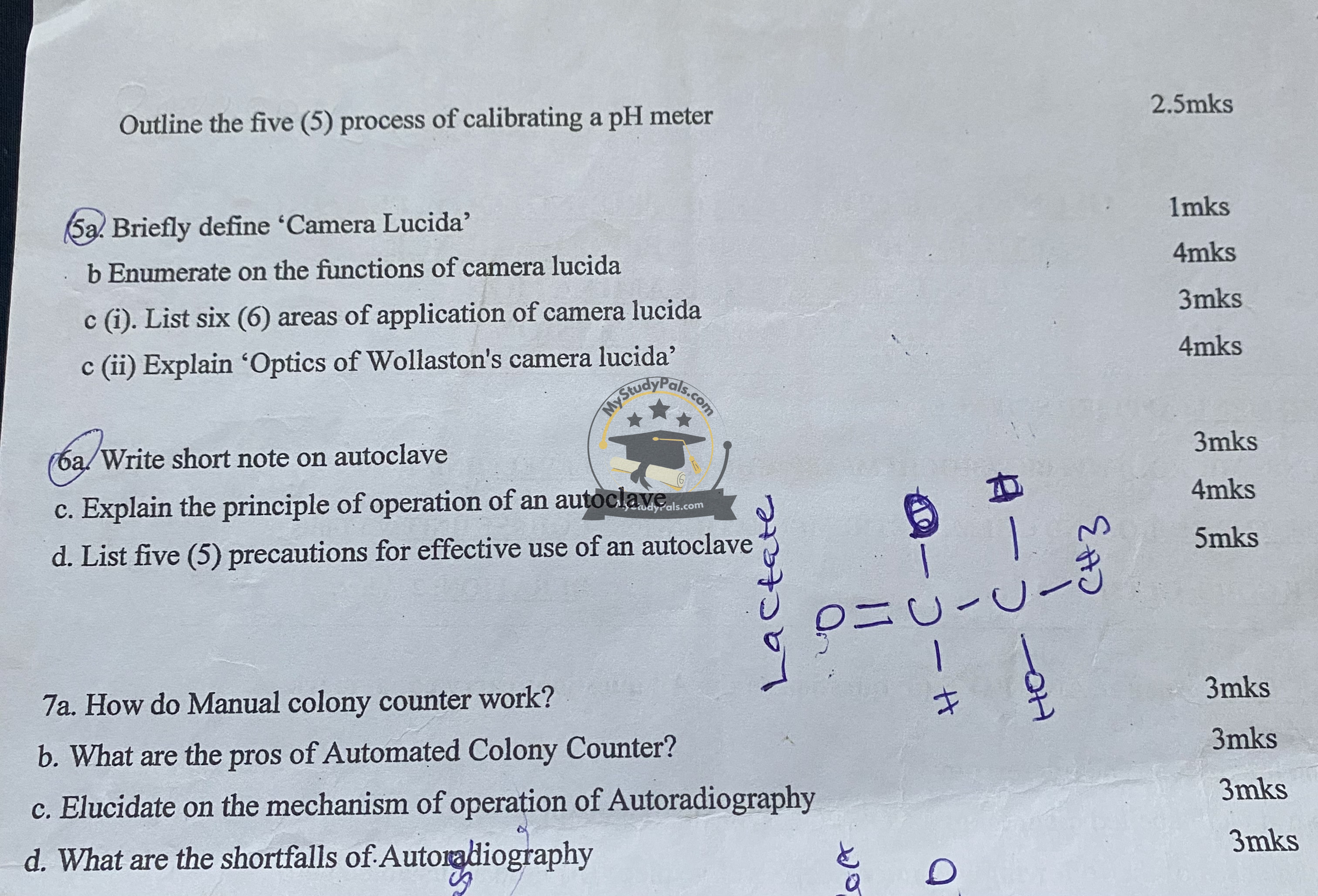
Outline the five (5) processes of calibrating a pH meter.
- Turn on the pH meter and allow it to warm up.
- Rinse the electrode with distilled water and dry it.
- Immerse the electrode in a standard buffer solution (e.g., pH 7.0).
- Adjust the meter to read the correct pH of the buffer.
- Repeat with additional buffers (e.g., pH 4.0 and 10.0) to ensure accuracy.
(a) Briefly define “Camera Lucida”.
- A Camera Lucida is an optical device that allows the user to superimpose an image of an object onto a drawing surface for accurate sketching.
(b) Enumerate the functions of Camera Lucida.
- Helps in detailed scientific drawing.
- Assists in scaling and proportion accuracy.
- Used in biological and medical illustrations.
- Facilitates accurate replication of microscopic observations.
(c)(i) List six (6) areas of application of Camera Lucida.
- Scientific research.
- Medical illustrations.
- Forensic sketching.
- Paleontology.
- Microscopy observations.
- Fine arts and architecture.
(c)(ii) Explain “Optics of Wollaston’s Camera Lucida”.
- Wollaston’s Camera Lucida uses a prism to reflect an image onto a surface, enabling the user to trace it while viewing both the object and the drawing simultaneously.
Question 6:
(a) Write a short note on autoclave.
- An autoclave is a device that uses high-pressure steam at high temperatures to sterilize equipment, media, and waste materials in laboratories and medical facilities.
(c) Explain the principle of operation of an autoclave.
- An autoclave works on the principle of steam sterilization. Steam under high pressure (121°C at 15 psi for 15-20 minutes) penetrates materials, killing bacteria, viruses, and spores by coagulating their proteins.
(d) List five (5) precautions for effective use of an autoclave.
- Do not overload the chamber.
- Ensure proper sealing of the door before operation.
- Use appropriate sterilization cycles for different materials.
- Allow proper cooling before opening to avoid burns.
- Regularly maintain and validate performance.
Question 7:
(a) How does a manual colony counter work?
- A manual colony counter uses a magnifying lens and light source to help a microbiologist visually count bacterial colonies on agar plates.
(b) What are the pros of an automated colony counter?
- Faster and more accurate counting.
- Reduces human error and fatigue.
- Provides digital records for documentation.
(c) Elucidate the mechanism of operation of autoradiography.
- Autoradiography detects radioactive isotopes in biological samples. A photographic film is placed over the sample, where radioactive emissions expose the film, creating an image of molecular distributions.
(d) What are the shortfalls of autoradiography?
- Requires handling of radioactive substances.
- Time-consuming exposure and development process.
- Limited sensitivity compared to modern imaging techniques.


